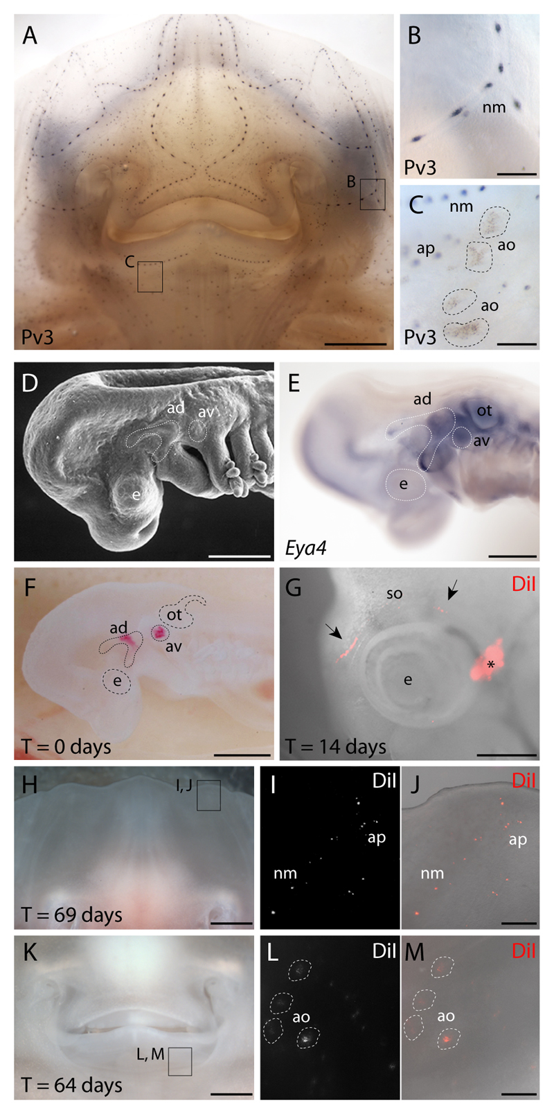
Fig. 1. Lateral line placodal origin of skate ampullary organs and neuromasts.
(A) Wholemount immunostaining for parvalbumin-3 (Pv3) in L. erinacea embryos reveals a cephalic network of (B) mechanosensory neuromasts and (C) electrosensory ampullary organs. In order to test the lateral line placodal origin of neuromasts and ampullary organs, we fate-mapped the anterodorsal or anteroventral lateral line placodes. These placodes were recognizable as (D) ectodermal thickenings caudal to the eye and dorsal to the mandibular and hyoid arches, respectively, which (E) express the transcription co-factor gene Eya4. (F) An embryo in which the anterodorsal and anteroventral lateral line placodes were focally labelled with DiI. (G) In an embryo in which the anterodorsal lateral line placode was labelled, DiI was observed at 14 days post-injection in the supraorbital lateral line primordium (black arrows), far from the original injection site (*). In embryos with DiI-labelled anterodorsal and/or anteroventral lateral line placodes, the distribution of DiI-positive cells at 60-70 days post-injection recapitulated the normal distribution of (H-J) cephalic neuromasts, ampullary pores and (K-M) ampullary organs. ad, anterodorsal lateral line placode; ao, ampullary organ; ap, ampullary tubule pore; av, anteroventral lateral line placode; e, eye; nm, neuromast; ot, otic vesicle; so, supraorbital lateral line primordium. Scale bars: A,H,K: 2.5 mm; B-C,J,M: 0.5 mm; D-E: 0.5 mm; G: 0.8 mm.

