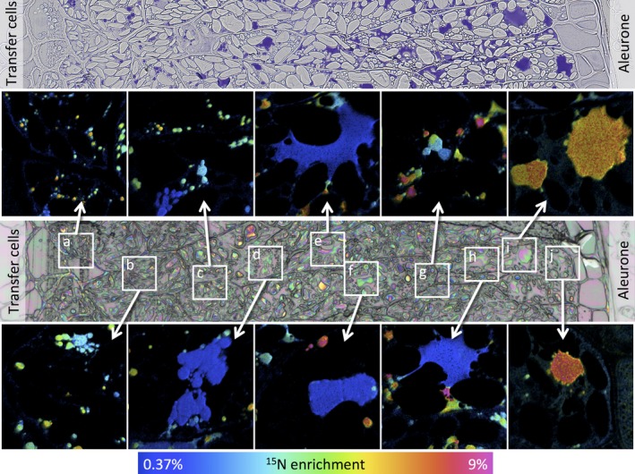
Figure 3.

Analysis of transect 1 of a wheat starchy endosperm taken from a developing caryopsis at 27 dpa, after feeding 15N glutamine at 20 dpa. The top panel shows a conventional light microscopy image after staining with toluidine blue to identify protein bodies. The central panel shows an optical image from a serial section which was used to select the areas where secondary ion NanoSIMS images were acquired at high lateral resolution. Areas a to j marked on this optical image are expanded in the boxes and enrichment with 15N is shown using a hue saturation intensity colour scale with the 15N enrichment shown in the scale at the bottom.
