
Figure 1.
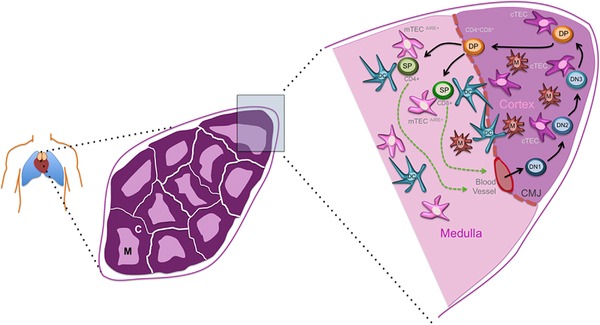
Thymus structure and development. Schematic representation of a human thymus. Left panel shows location of the thymus, at the midline above the heart. Middle panel shows representation of a section through a young thymus, indicating the thymic cortex (c) and medulla (m). Right panel shows detail of stromal cells (thymic epithelial cells, TECs; dendritic cells, macrophages, and blood vessels.) Note that mesenchymal cells and the vascular network are omitted for clarity, although the mesenchymal capsule bounding the thymus is shown. Hematopoietic progenitors enter the thymus at the junction between cortex and medulla. Commitment to the T‐cell lineage and differentiation as far as the CD4+CD8+ ‘double positive’ (DP) stage of development occurs in the cortex. Thymocytes that successfully undergo positive selection can then enter the medulla, which is the site of central tolerance induction. CD4+ and CD8+ single positive (SP) T cells exit the thymus from the medulla (see 2, 3, 4, 5, 6, 7). DN, CD4−CD8− ‘double negative’ thymocytes.