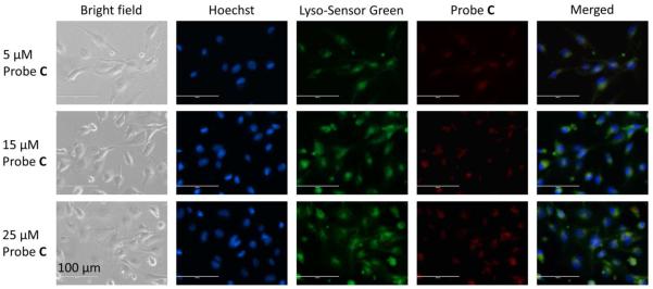
Figure 5.
Fluorescence images of HUVEC-C cells incubated with 5 μM, 15 μM, or 25 μM fluorescent probe C. HUVEC-C cells were incubated with fluorescent probe C for 2 h, post serum starvation (2 h) and imaged for co-localization with 1 μM LysoSensor Green and (1 μg·mL−1) Hoechst 33342 stains. Images were acquired using the inverted fluorescence microscope (AMF-4306, EVOSfl, AMG) at 40× magnification, scale bars = 100 μm.

