Fig. 2.
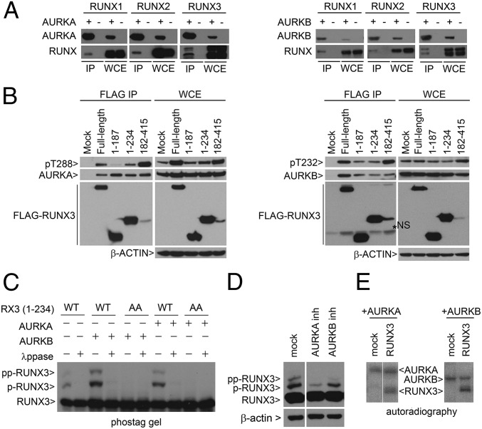
RUNX proteins interact with Aurora kinases A and B. (A) HEK293T cells were cotransfected with various combinations of RUNX1, RUNX2, RUNX3, Flag-AURKA, and GFP-AURKB expression vectors. Immunoprecipitation (IP) was performed using anti-Flag M2 beads to enrich for AURKA (Left) and GFP affinity beads to enrich for AURKB (Right), respectively. RUNX proteins were detected with anti–pan-RUNX antibody. WCE, whole cell extract. (B) COS7 cells were transfected with Flag-RUNX3 truncation constructs and GST-AURKA or AURKB expression constructs. Flag-RUNX3 constructs were precipitated by M2 beads. Bound AURKA (Left) or AURKB (Right) proteins were detected by immunoblot. (C) WT RUNX3 (amino acids 1–234) or double mutant AA (T14A/T173A) were transfected into COS7 with AURKA or AURKB expression plasmids. λppase, lambda phosphatase. Flag antibody was used to detect RUNX3 proteins. RUNX3, p-RUNX3, and pp-RUNX3 refer to the different degrees of phosphorylation. Absence of p refers to unphosphorylated RUNX3. (D) COS7 cells were transfected with Flag-RUNX3 (amino acids 1–234). Cells were treated with nocodazole, followed by inhibitors (inh) of AURKA (MLN8237, 5 µM) and AURKB (ZM4473439, 2 µM) kinase inhibitors for 4 h, and harvested by mitotic shake-off. (E) In vitro kinase reaction of recombinant RUNX3 with AURKA or AURKB in the presence of [γ32P]ATP. Proteins were resolved by SDS/PAGE and visualized by autoradiography.

