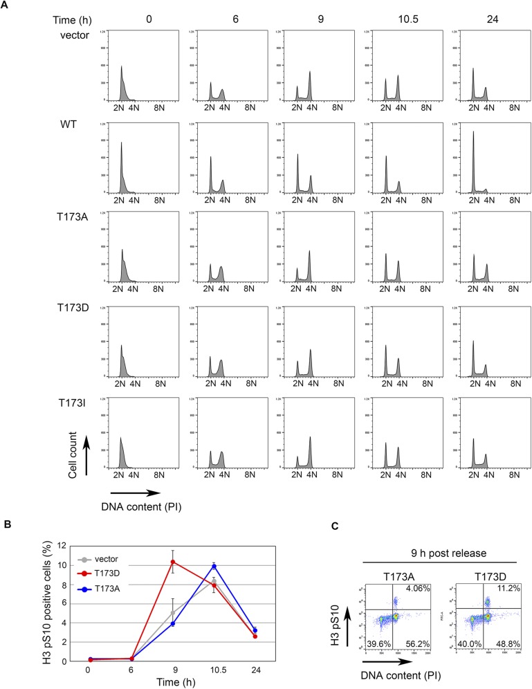
Fig. S4.
Flow cytometric analysis of cell cycle progression of pRetro-flag-RUNX3 stable HeLa Tet-On cell lines after release from thymidine block. (A) Histogram showing cell cycle profiles of the indicated p-RetroX-Flag-RUNX3 cell constructs at various time points after release from thymidine block. RUNX3 expression was induced with 0.5 µg/mL of doxycycline 16 h before release from block. Propidium iodide (PI) was used to quantify DNA content and the histogram of PI staining was plotted using FlowJo software. At time 0 h, cells were synchronized at G1/S phase with 2N DNA content. By 24 h, all cell lines except for WT RUNX3 have entered the next cell cycle. (B) Levels of mitotic cells of empty vector, T173A, and T173D cells in A were quantified by phospho-Histone 3 (Ser 10) (H3 pS10) staining. (C) Density plots of T173A and T173D at 9-h time point from B. Percentages of G2 and M subpopulations are indicated by elevated PI and H3 pS10 staining, respectively.

