Fig. 1.
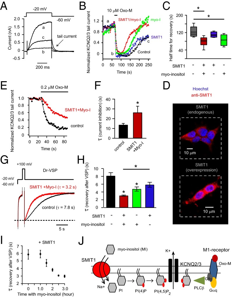
Overexpression of SMIT1 and incubation with myo-inositol slow inhibition and speed recovery of KCNQ2/3 current after PI(4,5)P2 depletion. (A) Representative current traces for KCNQ2/3 channels before (a), immediately after (b) 10 μM Oxo-M application, and during recovery (c). Arrow indicates the tail current. (B) Combinatorial treatments of SMIT1 overexpression with or without 100 μM myo-inositol on the KCNQ2/3 tail current recovery after M1 receptor activation. (C) Summary of the data in B, showing the half time for current recovery (n = 4–6). (D) Representative confocal immunocytochemistry images showing the expression of SMIT1 protein (red) in tsA201 cells, with counterstaining for nuclei (blue). (E) Time courses of inhibition of KCNQ2/3 current after applying low concentrations of Oxo-M (0.2 μM), comparing control cells with cells transfected with SMIT1 plus myo-inositol. (F) Summary of the data in D, shown as the exponential time constant τ of the current inhibition (n = 4–5). *P < 0.05. (G) Current traces, showing the inhibition and recovery of KCNQ2/3 current after activation of Dr-VSP. (H) Summary of the data in F, illustrating the effects of combinatorial treatments of SMIT1 overexpression with or without 100 μM myo-inositol on the time constants of KCNQ2/3 current recovery after Dr-VSP activation (n = 4–13). Means ± SEM, *P < 0.05. (I) Effects of different durations of 100 μM myo-inositol preincubation on the τ of KCNQ2/3 current recovery after Dr-VSP activation. (J) Cartoon showing the hypothesis that myo-inositol entry through SMIT1 raises the intracellular levels of phosphoinositides.

