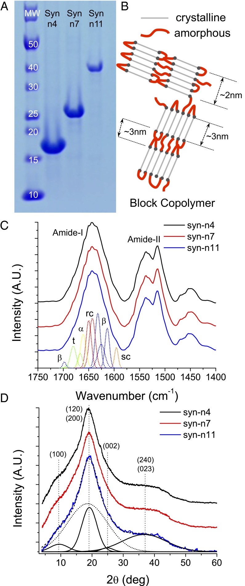
Fig. 3.
(A) SDS/PAGE showing the sizes of the synthetic proteins with n = 4, n = 7, and n = 11. (B) Cartoon representation of the segmented polymer architecture of assembled polypeptides containing ordered β-sheet crystals and amorphous Gly-rich regions. Amorphous and crystalline are colored in green and red, respectively. The (C) FTIR and (D) XRD spectra for all three samples are shown. α, α-helix, β, β-sheet; MW, molecular weight; rc, random coil; sc, side chain; t, turn.

