Figure 3.
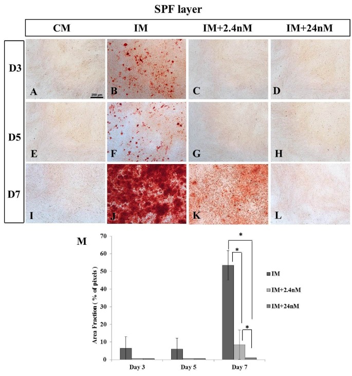
Inhibition of calcium deposition in SPF-Layer (Lohmann LSL) MSCs undergoing osteogenic differentiation in the presence of high and low concentrations of 1,25(OH)2D3 in vitro. Mineralization was observed at all time points in cells undergoing osteogenic induction in the absence of exogenous 1,25(OH)2D3 (B, F, J). No mineralization is observed at Day 3 or 5 in the presence of 2.4nM, (C, G) or 24nM (D, H) 1,25(OH)2D3. (K) Only small foci of mineralization are observed in low 2.4nM 1,25(OH)2D3 at Day 7 of osteogenic differentiation A, E, I) MSCs grown in the absence of osteogenic inducing factors show no mineralization on any day examined. M) Quantitative analysis of calcium deposition expressed as area fraction (% of pixels). Values are mean ± SEM (n = 4 on all days examined). Comparisons marked with an asterisk (*) are significantly different (p < 0.05). Scale bar = 200μm. Abbreviations: CM, control media containing no exogenous 1,25(OH)2D3; IM, induction media; IM+2.4nM, induction media containing 2.4nM 1,25(OH)2D3; IM+24nM, induction media containing 24nM 1,25(OH)2D3; D, Day.

