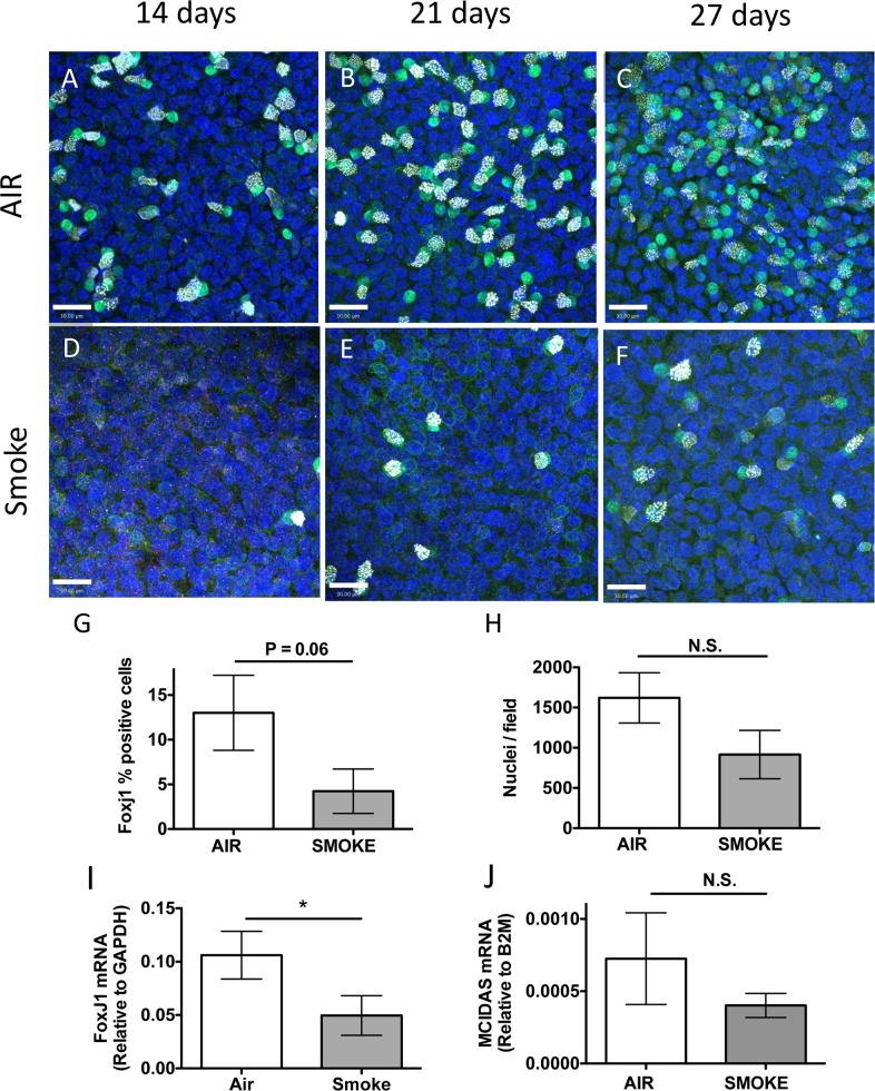
Fig 4. Whole cigarette smoke exposure inhibits ciliated cell differentiation.
(A–F) Representative confocal micrographs of human airway epithelial cells treated with Air (A, B, C) or Smoke (D, E, F) for 14 d (A, D), 21 d (B, E) or 27 d (C, F) during differentiation using ALI conditions and stained for FoxJ1 (green), acetylated-tubulin (white) or nuclei (blue). Scale bar = 30 μm. (G) Quantification of FoxJ1 positive cells treated with Air (white bars) or Smoke (grey bars) for 27 d during differentiation in ALI conditions. N = 3 different lung donors, * P = 0.06, two-tailed Student’s t test. (H) Quantitation of the average number of nuclei / microscopic field in cells treated with Air (white bar) or WCS (grey bar) for 27 days during differentiation. N = 3 different lung donors, N.S., not significant, two-tailed Student’s t-test. (I) Duplex qRT-PCR of FoxJ1 mRNA relative to GAPDH mRNA in cells treated with Air or Smoke for 27 d during differentiation using ALI conditions. N = 8 different lung donors, * P<0.05, two-tailed Student’s t-test. (J) Duplex qRT-PCR of MCIDAS mRNA relative to B2M in human airway epithelial cells treated with Air (white bars) or Smoke (grey bars) for 27 d of differentiation in ALI conditions. N = 8 different lung donors, N.S., not significant, two-tailed Student’s t-test.

