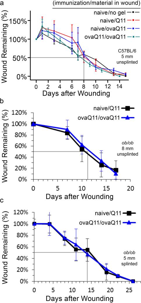
Figure 2. The rate of wound healing is unaffected by high antibody titers against the scaffold.
Wound closure rates are shown for 3 different models of increasingly delayed healing. Gray lines indicate approximate times to 50% healing. Naming convention: “(immunization material/scaffold material)” (a) The 5 mm unsplinted C57BL/6 model demonstrated no significant difference in wound healing rate between (ovaQ11/ovaQ11), (naïve/Q11), (naïve/PBS), or (naïve/ovaQ11). n= 3 mice per group, except for ovaQ11/ovaQ11 for which n=2 because one mouse died during anesthesia. (b) 8 mm unsplinted ob/ob model demonstrated no significant difference in wound healing rates between (naïve/Q11) and (ovaQ11/ovaQ11). n= 9 mice per group. (c) Splinted 5 mm ob/ob wound model demonstrated no significant difference in wound healing rates between (naïve/Q11) and (ovaQ11/ovaQ11), n=6 mice per group.

