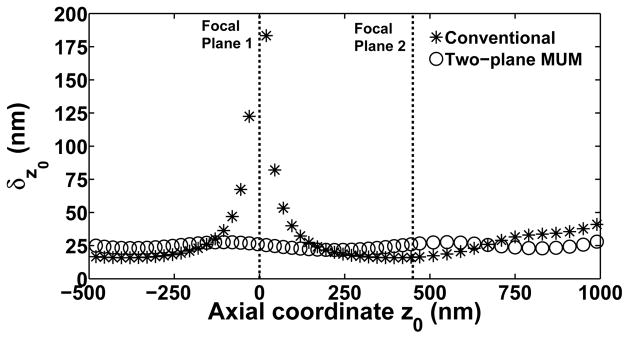
Fig. 9.
Limits of the axial coordinate estimation accuracy for a conventional microscopy setup and a two-plane MUM setup. The limit of accuracy δz0 for the estimation of the z0 coordinate of an out-of-focus and stationary molecule from a conventional image (*) or a pair of two-plane MUM images (○) is plotted as a function of the coordinate z0. Focal planes 1 and 2 of the MUM setup are denoted by vertical dashed lines, and are located at z0 = 0 nm and z0 = 450 nm, respectively. The value of z0 is specified with respect to focal plane 1, which coincides with the focal plane of the conventional setup. In both setups, an image consists of an 11×11-pixel region, acquired using a CCD detector with a pixel size of 13 μm × 13 μm and readout noise with mean η0 = 0 electrons and standard deviation σ0 = 6 electrons at each pixel. The image of the molecule is described by the Born and Wolf image function. For the conventional setup, the molecule is laterally positioned such that the center of its image is located at 5.05 pixels in the x direction and 5.25 pixels in the y direction. Photons are detected from the molecule at a rate of Λ0 = 10000 photons/s over an acquisition time of t − t0 = 0.1 s, such that Λ0 · (t − t0) = 1000 photons are on average detected from the molecule over the detector plane (and 952.3 photons are on average detected from the molecule over a given image when the molecule is in focus). A background detection rate of is assumed, along with a uniform background spatial distribution of fb(x, y) = 1/20449 μm−2, such that there is a background level of β0 = 20 photoelectrons per pixel over the 11×11-pixel image. Photons of wavelength λ = 520 nm are collected by an objective lens with magnification M = 100 and numerical aperture na = 1.4. The refractive index of the immersion medium is n = 1.518. For the MUM setup, the collected molecule and background photons are split equally between the images corresponding to the two focal planes. Therefore, the photon detection rate is Λ0 = 5000 photons/s per image, and the background detection rate is per image. For the image corresponding to focal plane 1, all other details are as given for the conventional setup. For the image corresponding to focal plane 2, the magnification is M = 98.18, calculated using a formula from [76] for a plane spacing of Δzf = 450 nm and a tube length of 160 mm. The lateral position of the image of the molecule and the background spatial distribution are accordingly adjusted based on this slightly smaller magnification.

