
Figure 6. Injection of PLGA-4a1 resulted in increased HEXIM1 expression and decreased metastasis.
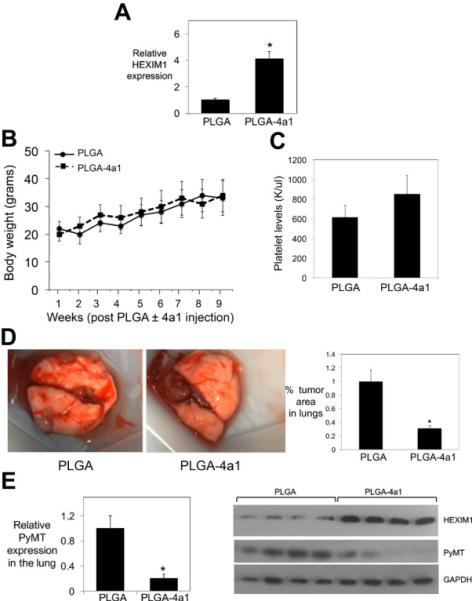
After the appearance of palpable mammary tumors in PyMT mice, PLGA or PLGA-4a1 (50 uM, 50 ul volume) were injected into the tumors every other week. Mammary glands, blood, and lungs were then collected. (A) HEXIM1 expression in mammary tumors of PLGA ± 4a1 treated mice were determined by Western blot analyses. (B) Body weights were monitored weekly as indicated. (C) Platelet levels were determined using the HEMAVET 950FS Multi-species Hematology System. (D) Left panel shows lungs from PLGA or PLGA-4a1 treated mice. Right panel shows quantification of tumor area in H&E stained lung tissue sections. (E) Western blot analyses of PyMT expression in the lungs of PLGA or PLGA-4a1 treated mice. In (A) and (E), expression of HEXIM1 and PyMT, respectively, are expressed relative to expression of GAPDH, a loading control. Panels represent 5 mice per group (PLGA ± 4a1). * p < 0.01 relative to PLGA treated cells.