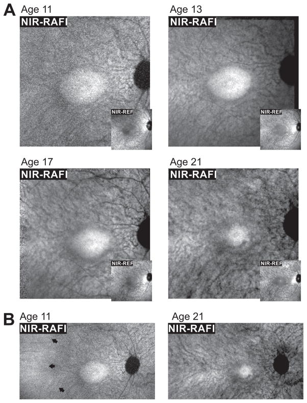
FIGURE 3.
Serial changes apparent using en face imaging with a decade of followup. (A) Near-infrared (NIR) excited reduced-illuminance autofluorescence imaging (RAFI, main panels) and reflectance (REF, insets) imaging of the macula in the patient with SPATA7 mutations at ages 11, 13, 17 and 21. A central homogeneous-appearing elliptical region of higher NIR-RAFI signal and lower NIR-REF signal is surrounded by a heterogeneous region displaying visibility of choroidal blood vessels. (B) Digital composites of NIR-RAFI showing a region of relatively greater homogeneity temporal to the macula (arrows) at age 11 that is not detectable by age 21. Each image is individually contrast stretched to allow visibility of features. Images shown in Panel A are 30°×30° and those in Panel B are 51°×29°.

