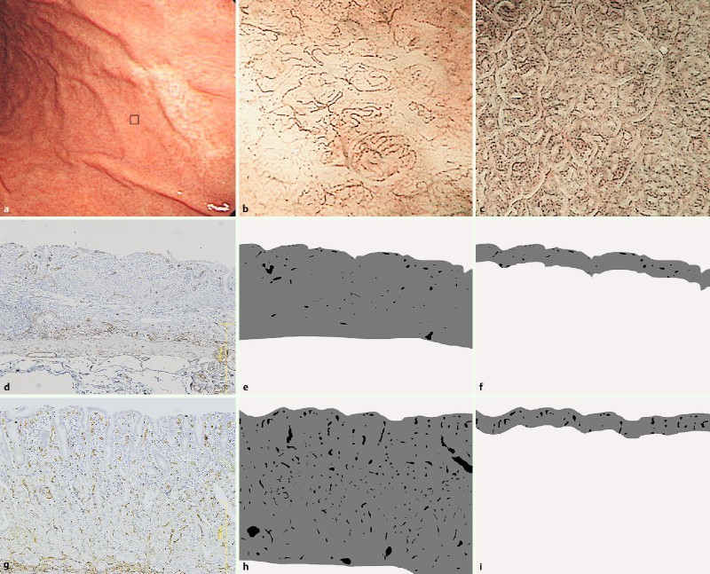
Fig. 2 a.

Discolored undifferentiated type gastric carcinoma in the middle corpus. b Microvessels were extracted from the ME-NBI image (white box in a indicates location of b). The NBI vascular density was 4.34 % in the carcinoma. c The NBI vascular density was 10.59 % in the surrounding uninvolved mucosa (black box in a indicates location of c). d Photomicrograph showing a CD34 immunostained tissue section from a carcinoma. e The histologically assessed vascular index was 1.49 % in the whole mucosal layer. f The histologically assessed vascular index was 2.56 % in the superficial mucosal layer (0 – 100 μm). g Photomicrograph showing a CD34 immunostained tissue section from the surrounding uninvolved mucosa. h The histologically assessed vascular density was 5.84 % in the whole mucosal layer. I The histologically assessed vascular density was 6.87 % in the superficial mucosal layer (0 – 100 μm).
