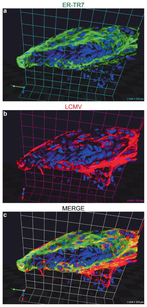
Fig. 1.
Localization of LCMV in the meninges following intracerebral inoculation. Sagittal brain vibratome sections obtained from day 6 LCMV-infected mice were stained with antibodies directed against ER-TR7 (green) and LCMV (red). ER-TR7 is a stromal cell marker that labels fibroblast-like cells. A representative 3D reconstruction of a meningeal blood vessel cross section is shown to illustrate the degree to which LCMV infects fibroblast-like cells that completely surround meningeal blood vessels. (Grid scale = 19.5 μm). Cell nuclei are shown in blue

