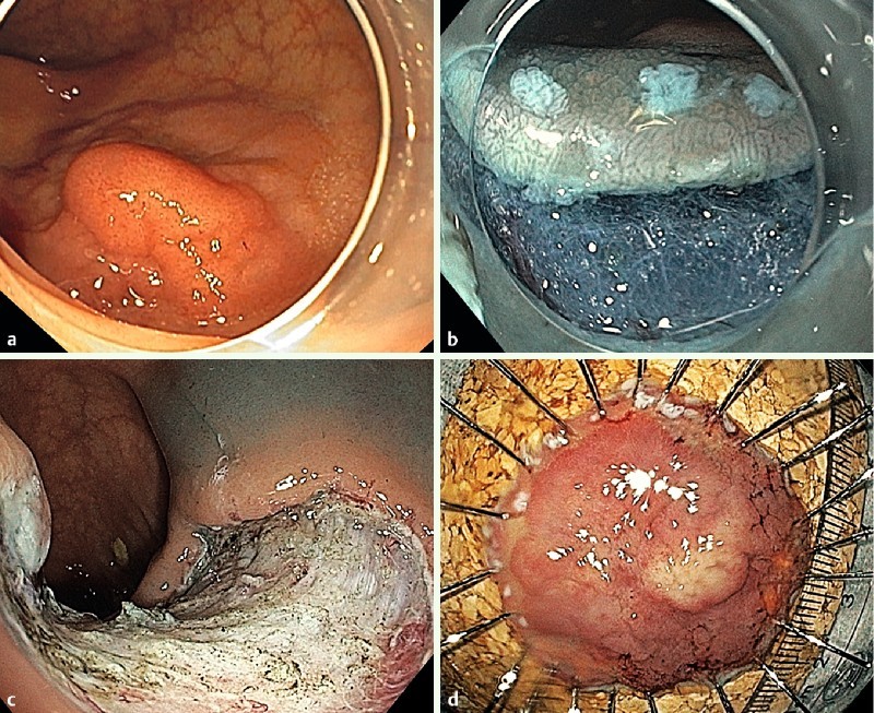
Fig. 1.

a ESD of a high grade IEN in the ascending colon. Aspect of the lesion (0-IIa/0-Is; LST-granular nodular). b Initial incision of the mucosal layer and submucosal dissection. Note the marking dots. c Aspect of the resection site. d Specimen pinned on corkboard.
