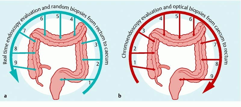
Fig. 1.

Two-step colonoscopy procedure a During the intubation phase of colonoscopy (from rectum to cecum) we performed high-definition endoscopic evaluation of the colon mucosa. All visible lesions were assessed for size and location but not biopsied. Random biopsies were taken from 9 predefined locations corresponding to every 10 cm of the colon, starting from the rectum and ending in the cecum. b During the withdrawal phase, all visible lesions were defined using chromoendoscopy. After HDE/chromoendosopy evaluation, each abnormal lesion was inspected using pCLE and targeted biopsies were taken whenever a suspicion of dysplasia in HDE/chromoendoscopy and/or pCLE occurred. All areas where the random biopsies had been obtained during the intubation phase were also inspected with pCLE during the withdrawal phase.
