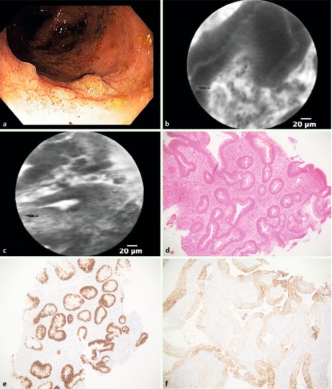
Fig. 3.

Low-grade dysplasia within visible lesion in the right colon. a Colonoscopy detected a visible lesion in the right colon. b and c pCLE showed elongated, irregular crypts with dark epithelium and fluorescein leakage. d Histopathology revealed epithelial atypia with cigar-shaped nuclei with pseudostratification, magnification 20 ×. e Immunohistochemistry showed stronger labeling for p53 in the dysplastic-appearing area than in surrounding crypts (20 ×), and f extensive labeling for cytokeratin 7 (40 ×).
