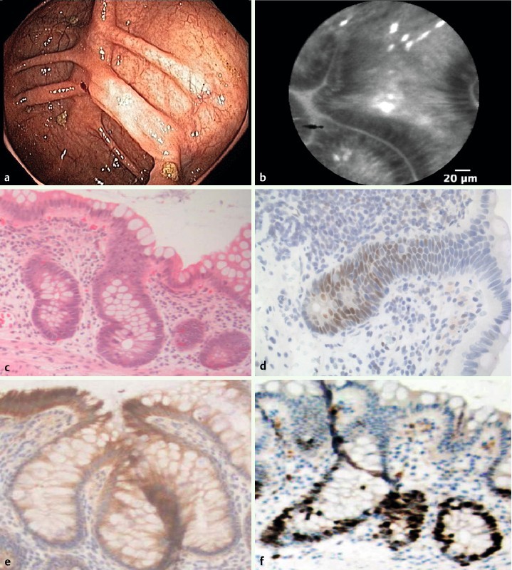
Fig. 5.

Epithelial atypia with increased proliferative activity but without the presence of dysplasia in the right colon mucosa. a Colonoscopy showed an inactive IBD without epithelial irregularities. b pCLE showed elongated and irregular crypts with dark epithelium. c Histopathology revealed epithelial atypia (40 ×). d Immunohistochemistry showed weak labeling for p53 and e P405S (40 ×). f Mib-1 staining showed increased proliferative activity (40 ×).
