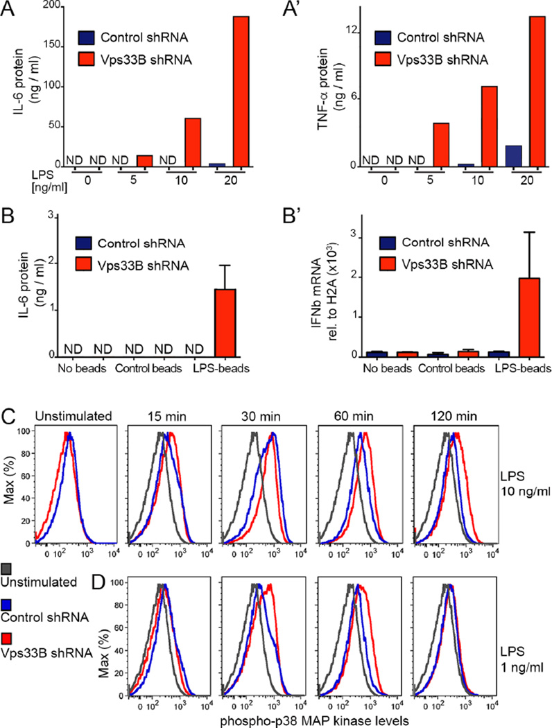
Figure 3. Vps33B silencing sensitizes macrophages to low LPS doses.
A) ELISA measurements of IL-6 (A) and TNF-α (A’) in the supernatants of Vps33B-deficient or control macrophages exposed to the indicated concentrations of LPS for 12 hours.
B) Vps33B-silenced or control macrophages were exposed to control beads or LPS-bound beads for 6 hours. Secreted IL-6 was measured by ELISA (B) or IFNβ transcript relative to Histone2A by qRT-PCR (B’).
(“ND” indicates not detected).
C,D) Flow cytometry analysis of phospho-p38 MAP kinase at indicated time points after stimulation with 10 ng/ml (C) or 10 ng/ml of LPS (D).
Data in this figure here are representative of 3 independent experiments. Please also see Figure S2.

