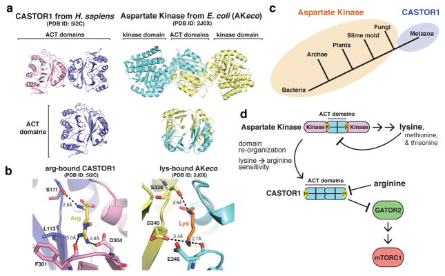
Figure 5. Insights into the evolution of arginine sensing by CASTOR1.
a, (Top) Ribbon view of human CASTOR1 dimer (pink and purple) and AKeco dimer (blue and yellow, PDB ID 2J0X). (Bottom) Ribbon view of the human CASTOR1 monomer (left) and regulatory domain from AKeco (right). b, Comparison of the arginine-binding pocket in human CASTOR1 with the lysine-binding pocket in AKeco. Arginine and lysine are shown in yellow and orange, respectively. Hydrogen bonds and salt-bridges are shown as black dashed lines. c, Phylogenetic distribution of Aspartate Kinase (orange) and CASTOR1 homologues (purple). d, Model of the evolution of CASTOR1 from the regulatory domain of an ancestral Aspartate Kinase.

