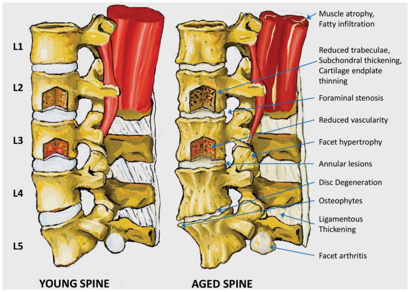
Figure 2. Gross features of the aging spine.
Young and aged lumbar spines are visually compared to illustrate the wide range of tissues and processes involved in aging of the spine. Muscle atrophy and fatty infiltration is evident at L1-2 in the aged spine. 25Similarly, a window into L3 depicts reduced vascularity and fewer capillaries reaching the endplate. Foraminal stenosis is shown at L2-3 (arrow), L3-4, and L4-5, and facet hypertrophy is evident at L3-4 (arrow) and L4-5. An annular lesion is present in the posterior portion of the L3-4 disc. Disc degeneration with evident loss of disc height and prominent anterior osteophytes occur at L4-5. Ligamentous thickening is indicated in the interspinous and supraspinous ligament; thickening of the ligamentum flavum occurs with aging but is not observable in the sagittal view. Finally, facet cartilage arthritis is revealed on the inferior facet of L5.

