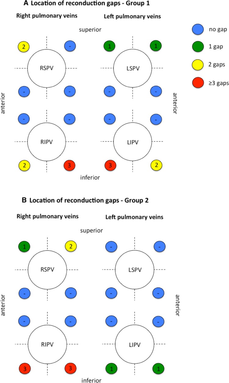
Fig. 3.
Location of electrical reconduction gaps. The figure depicts the number and location of reconduction gaps identified during re-ablation procedures. a Findings for group 1 (bonus-freeze protocol), b findings for group 2 (no bonus-freeze protocol). Septal and lateral pulmonary vein ostia are divided into four segments (antero-superior, antero-inferior, postero-superior, postero-inferior). Numbers express reconduction gaps found for each segment. No gaps were found along the carina between the ipsilateral pulmonary veins. Data for a single left common pulmonary vein is not shown (each group n = 1 single left common pulmonary vein, each with one gap. RSPV right superior pulmonary vein, RIPV right inferior pulmonary vein, LSPV left superior pulmonary vein, LIPV left inferior pulmonary vein)

