Figure 6.
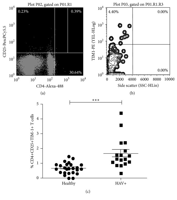
HAV infection leads to an increase in the relative proportion of TIM-1 receptor on CD4+CD25+ TLs. PBLCs from HAV+ pediatric patients were stained with anti-CD4-Alexa 488, anti-CD25-PerCP and anti-TIM1-PE antibodies and evaluated by using flow cytometry. (a) Representative dot plot of CD4+CD25+ staining and selection of the right upper quadrant. (b) Representative dot plot of TIM-1 versus Side Scatter linked to the right upper quadrant on (a), which represents TIM-1 percentage on CD4+CD25+ cells. (c) The results are displayed as the percentage of TIM-1+CD4+CD25+ T cells. The medians and standard deviations from 25 healthy donors and 17 HAV-infected pediatric patients are presented. Nonparametric Mann-Whitney U test for comparison between groups was used to calculate statistical significance. P < 0.05 was considered statically significant. ∗∗∗ P < 0.0001.

