FIGURE 3.
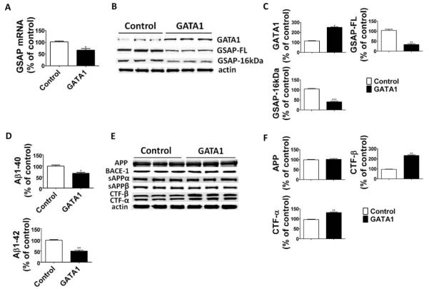
GATA1 overexpression decreases γ-secretase activating protein (GSAP) levels and reduces Aβ formation. N2A-APPswe cells were transfected overnight with GATA1 cDNA or empty vector (control). Supernatant and cell lysates were collected for analysis. (A) Relative mRNA levels for GSAP in cells transfected with GATA1 cDNA or empty vector (n = 3 control, and n = 3 GATA1; *p = 0.01; unpaired t test). (B) Representative Western blot analysis of GATA1, full-length GSAP (GSAP-FL), and GSAP-16kDa in cells transfected with GATA1 cDNA or empty vector. (C) Densitometric analyses of the immunoreactivities to the antibodies shown in B (n = 3 control, n = 3 GATA1; *p < 0.0001, **p = 0.0006, ***p = 0.0001; unpaired t test). (D) Aβ1–40 and Aβ1–42 levels in conditioned medium from cells transfected with GATA1 cDNA or empty vector were measured by sandwich enzyme-linked immunosorbent assay (n = 3 control, n = 3 GATA1; *p = 0.003, **p = 0.0002; unpaired t test). (E) Representative Western blot analysis of Aβ precursor protein (APP), BACE-1, sAPPα, sAPPβ, carboxyl-terminal fragment (CTF)-β, and CTF-α in cells transfected with GATA1 cDNA or empty vector. (F) Densitometric analyses of the immunoreactivities to the antibodies shown in E (n = 3 control, n = 3 GATA1; **p = 0.0008; unpaired t test). Values represent mean ± standard error of the mean. Results are presented as percentage change from the mean value of the control group.

