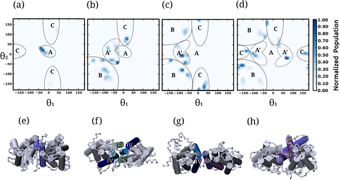
Figure 2. Relative orientations of the receptors in the dimer state.
Normalized population of the relative orientations of the two receptors in the dimer regime defined by the angles θ1 and θ2 (see Methods for details). The populations were averaged over the dimer regime for simulations in (a) POPC bilayers and POPC/cholesterol bilayers with (b) 9 (c) 30 and (d) 50% cholesterol concentrations. The relative orientations can be broadly mapped to four conformations (A, B, C and A′) that are marked in panels (a–d). The four conformations correspond to two sites at the receptor: site 1 comprising of transmembrane helices I and II and site 2 comprising of transmembrane helices IV, V and VI. (e) Conformation A corresponds to a non-flexible homo-interface with only a single helix from site 1 (transmembrane helix I). (f) Conformation B corresponds to homo-interfaces at site 2. (g) Conformation C corresponds to hetero-interfaces comprising of transmembrane helices from sites 1 and 2. (h) Conformation A′ corresponds to a flexible interface at site 1.

