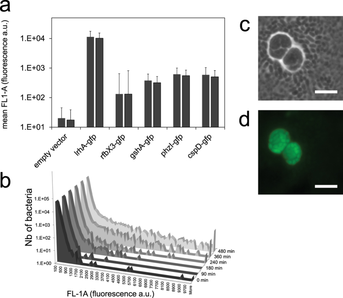
Figure 6. Expression of symplasmata-associated genes in Pantoea eucalypti as measured by GFP reporter fusions.
(a,b) Intensity of GFP fluorescence measured by flow cytometry in individual Pe299R bacteria carrying a reporter plasmid. (a) Histograms show the mean GFP fluorescence intensity (FL1-A) in individual bacteria after 8 hours of growth in M9G liquid cultures, expressed as arbitrary units and calculated from a sample of 10,000 cells; bars show results from two independent experiments; error bar indicates 1 SD. (b) Count histograms of Pe299R bacteria carrying the pRfbX3-gfp plasmid; we show the distribution of 100,000 cells after 0, 1.5, 4, 6 and 8 hours of incubation. (c,d) Micrographs of Pe299R (pRfbX3-gfp) after 21 hours of growth on the surface of M9G agarose. A symplasmatum surrounded by single cells is shown. Phase contrast (c) and GFP fluorescence with pseudo-color green (d) are shown. Bar is 5 μm.

