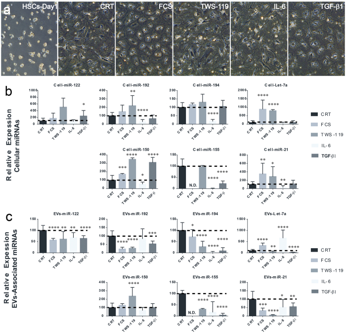
Figure 9. Modulation of miRNA expression and secretion in activated rat hepatic stellate cells.
Following activation, rat HSCs were cultured for 24 hours in serum free medium before stimulation with either FCS or the GSK-3β inhibitor TWS-119 or cytokines (IL-6 and TGF-β1) for 72 hours. (a) Effect of treatments on HSCs morphology was assessed via contrast phase microscopy. Expression profiling of selected miRNAs was analyzed by miQPCR in either (b) cellular or (c) vesicles-associated RNAs. The values of unstimulated controls HSCs (CRT) were set arbitrarily to 100. Statistical analysis was performed by unpaired T-test of control group (CRT; n = 12) versus individual treatment groups (n = 9 to 12) for each miRNA. Data are represented as average ± standard deviation. *P ≤ 0.05; **P ≤ 0.01; ***P ≤ 0.0005; and ****P ≤ 0.0001. N.D. Non-detectable.

