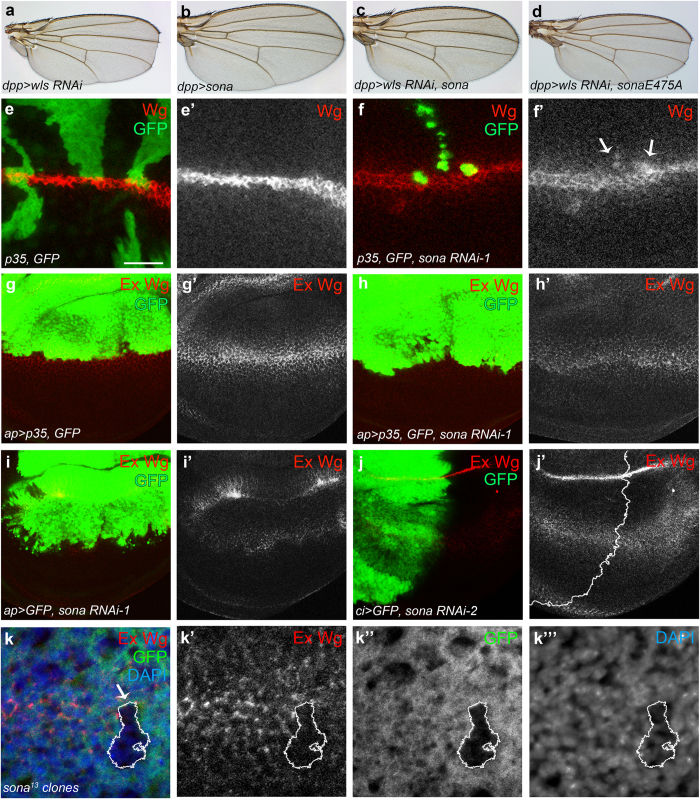
Figure 6. Loss of Sona increases the level of intracellular but decreases extracellular Wg.
(a–d) Notched wing phenotype of dpp > wls RNAi flies (a) was rescued by expression of sona (c) but not sonaE475 protease mutant form (d). dpp > sona wings had no phenotype (b). (e,f) Intracellular Wg in sona RNAi clones. hs-Flp, P[Actin > yellow > Gal4; w + ]/+; UAS-p35, UAS-GFP/+ wing disc as a control (e). sona RNAi-1 clones in hs-Flp, P[Actin > yellow > Gal4; w +]/+; UAS-p35, UAS-GFP/+; UAS-sona RNAi-1/+ (f). Arrows indicate the sona RNAi-1 clones with the increased level of intracellular Wg (f′). (g-i) Changes in the level of extracellular Wg by sona RNAi expression. UAS-p35, UAS-GFP/apterous (ap)-Gal4; +/TM6 Tb as a control (g), UAS-p35, UAS-GFP/ap-Gal4; UAS-sona RNAi/+ (h), and UAS-GFP/ap-Gal4; UAS-sona RNAi/+ (i). (j) Decrease in the extracellular level of Wg in the anterior region of ci > sona RNAi-2 discs. The control is shown in Supplementary Fig. S11. (k) Decrease in the level of extracellular Wg in sona13 clones near the DV boundary. Scale bar: (e,f) 40 μm; (g–j) 60 μm; (k) 9.5 μm.

