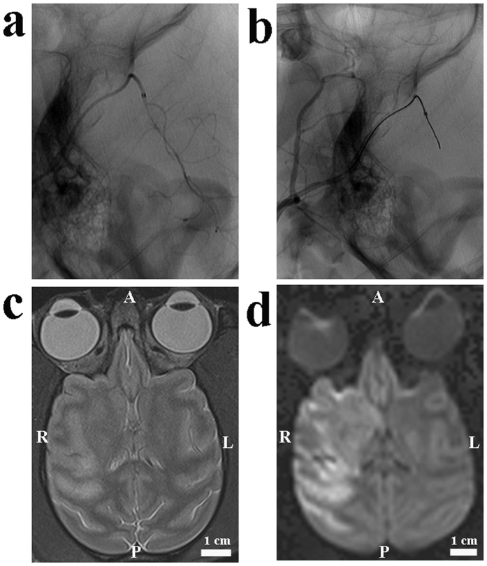
Figure 2. Images from an animal undergoing ischemia through the micro-catheter method.
(a) A lateral pre-occlusion angiogram demonstrated normal MCA, its divisions and the position of the micro-catheter. (b) A lateral post-occlusion angiogram illustrated the perfusion deficit in the M1 main trunk. (c) There was a prolonged T2, indicating early vaso-genic edema. (d) Diffuse hyper-intensity on DWI scans indicated ischemic lesions. A: anterior; P: posterior; R: right; L: left.

