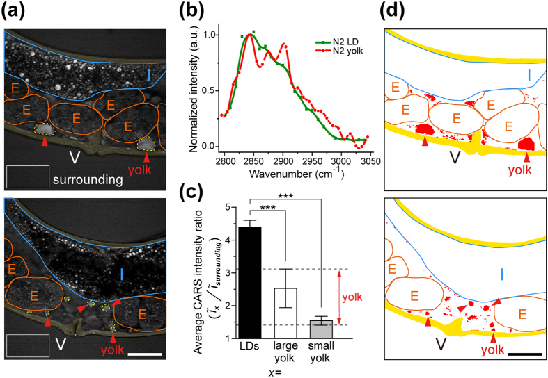
Figure 2. Analysis of yolk lipoprotein accumulation in the pseudocoelomic cavity of one-day-adult (1D-Ad) wild-type (N2) worms.
(a) The CARS image of 1D-Ad N2 worms containing large (upper image) and small (lower image) yolk lipoprotein accumulation (red arrows) in their pseudocoelomic cavities. The white rectangles are selected for calculating the average CARS intensity of surrounding buffer. Yellow region stands for the skin-like hypodermal cells, E stands for embryo, I stands for intestine, and V stands for vulva. Scale bar = 30 μm. (b) The CARS spectra of yolk lipoprotein accumulation obtained from pseudocoelomic cavity and the lipid droplet (LD) obtained from the skin-like hypodermal cell in 1D-Ad N2 worms. (c) The average CARS intensity ratios between LDs/surrounding buffer and between (large and small) yolk lipoprotein accumulation/surrounding buffer. A threshold of 1.47–3.12 (in red color) was applied to the CARS image to enhance the boundary of yolk lipoprotein accumulation. Error bars represent the standard deviation. (P-value: *p < 0.05, **p < 0.01, ***p < 0.001) (d) After excluding the internal organs/tissues (such as intestine, oocyte/embryo, and body wall) and applying the threshold, the pixels with intensity ratio between 1.47–3.12 are converted into red color, and pixels with intensity ratio <1.47 and >3.12 are converted into white color. The boundary of yolk lipoprotein accumulation can be easily distinguished (indicated by red arrows).

