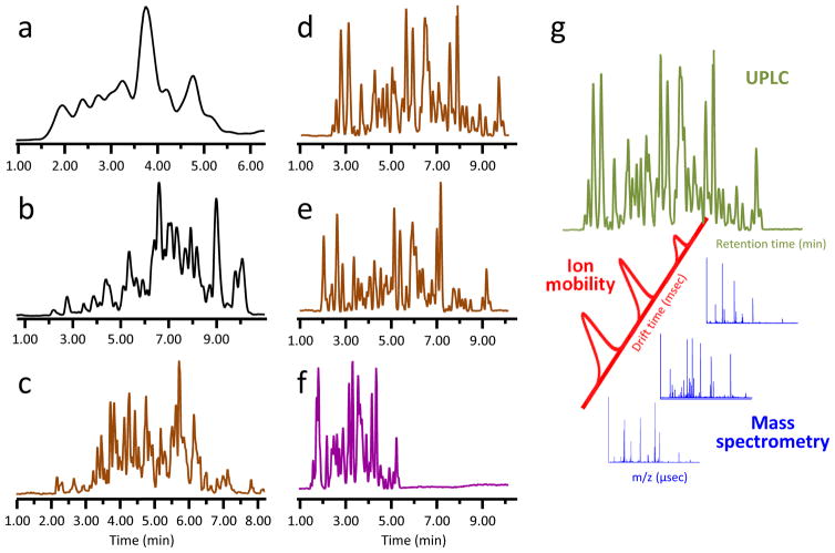
Figure 2.
Improving HX MS chromatographic separations at 0 °C. Panels (a–c) are taken from Ref. (132), with permission, and correspond to separation of a peptic digestion of a 52 kD protein with 20, 3.5, and 1.7 μm diameter particles, respectively. Panels (d–g) are separations of peptic digestion of monoclonal antibody (IgG) using a 1×50 mm HSS T3 1.8 μm column and a 5–35% water:acetonitrile gradient with changes to gradient time, flow rate, and backpressure: (d) 12 minute separation, 65 μL/min., backpressure 8000 psi; (e) 12 minute separation, 100 μL/min., backpressure 12000 psi; (f) 6 minute separation, 100 μL/min., backpressure 12000 psi, with addition of ion mobility separation. Separation with >2 μm particles (black traces, panels a,b) is inferior to separations with sub-2 μm particles (brown traces, panels c,d,e). Separation with both chromatography and ion mobility (purple trace, panel f) greatly enhances peak capacity. (g) Ion mobility separations require milliseconds and fit nicely between the time scale of liquid chromatography (minutes) and that of time-of-flight MS detection (microseconds).

