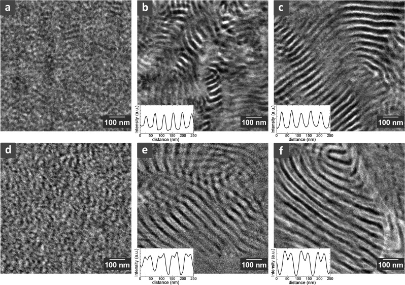
Figure 2.
Morphologies of dry and hydrated S-SES membranes by cryogenic scanning transmission electron microscopy (cryo-STEM). All the samples were unstained. (a–c) STEM images of dry S-SESA, S-SESB, and S-SESC, respectively. Typical line scan results through the alternating lamellae in S-SESB and S-SESC are shown as insets of (b) and (c), respectively. (d–f) Cryo-STEM images of hydrated S-SESA, S-SESB, and S-SESC, respectively. Typical line scans of hydrated S-SESB and S-SESC are shown as insets of (e) and (f), respectively. The large white feature on the righthand side of (f) is the lacey carbon support.

