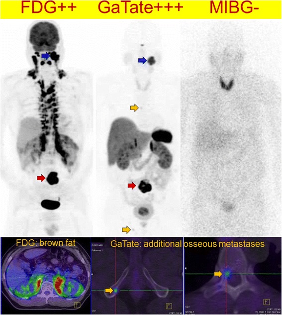
Fig. 7.

Patients with SDHB-related metastatic PGL showing extensive brown adipose tissue above and below the diaphragm on F-18 FDG PET/CT due to high circulating catecholamines (serum normetadrenaline 18 000 pmol/L, normal <900). F-18 FDG MIP (left) and axial fused PET/CT (bottom) and MIBG (right). The peri-nephric and, to a lesser extent central para-vertebral distribution is characteristic of PCC/PGL-related brown fat activation. Ga-68 DOTATATE (middle) clearly demonstrated an Organ of Zuckerkandl primary and several osseous metastases (botttom middle and bottom right), and is not subject to this physiologic artefact. As well as masking potential primary tumour sites along the sympathetic chain, the “sink effect” in brown fat limits availability of tracer for uptake in tumour sites
