Figure 5.
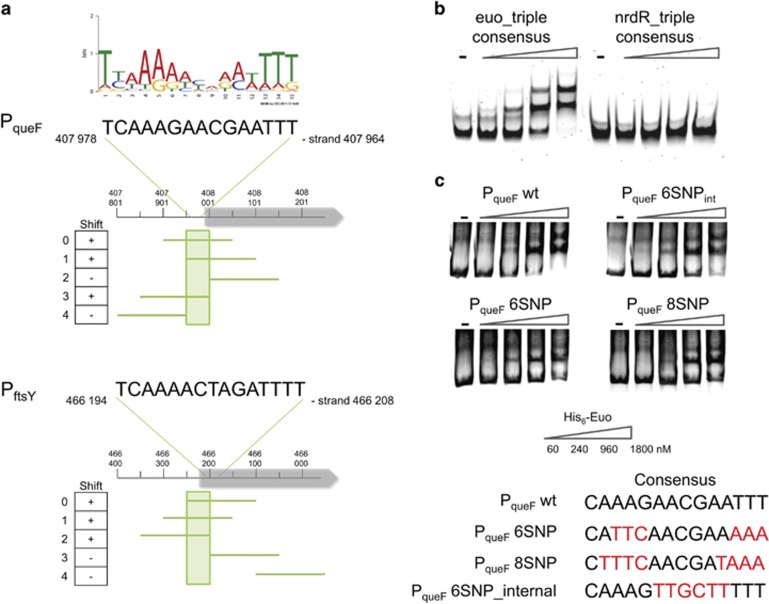
Euo consensus binding site analysis. (a) Identification of 50-bp region which includes the consensus and is necessary for the binding of His6-Euo. PCR probes shifted by 50 bp were designed for the PqueF and PftsY promoters and all fragments were used for in vitro binding assay (EMSA) with His6-Euo. A total of 80 ng of DNA fragments were incubated in the absence or in the presence of an increasing concentration of His6-Euo, protein–DNA complexes were detected using GelRed. Results are presented in the left column. + or − indicates whether or not shifted bands were observed. (b) EMSA analysis of His6-Euo binding to the Euo-triple consensus amplified with a specific primer coupled to Cy5. As a negative control, we used the triple repeat of the predicted NrdR consensus binding site (see Figure 4b). (c) Mutations in the consensus poorly affect the binding of His6-Euo tested by EMSA (GelRed detection, see Supplementary Information). His6-Euo concentrations used in the gel shift assay are indicated in (c).

