Figure 1.
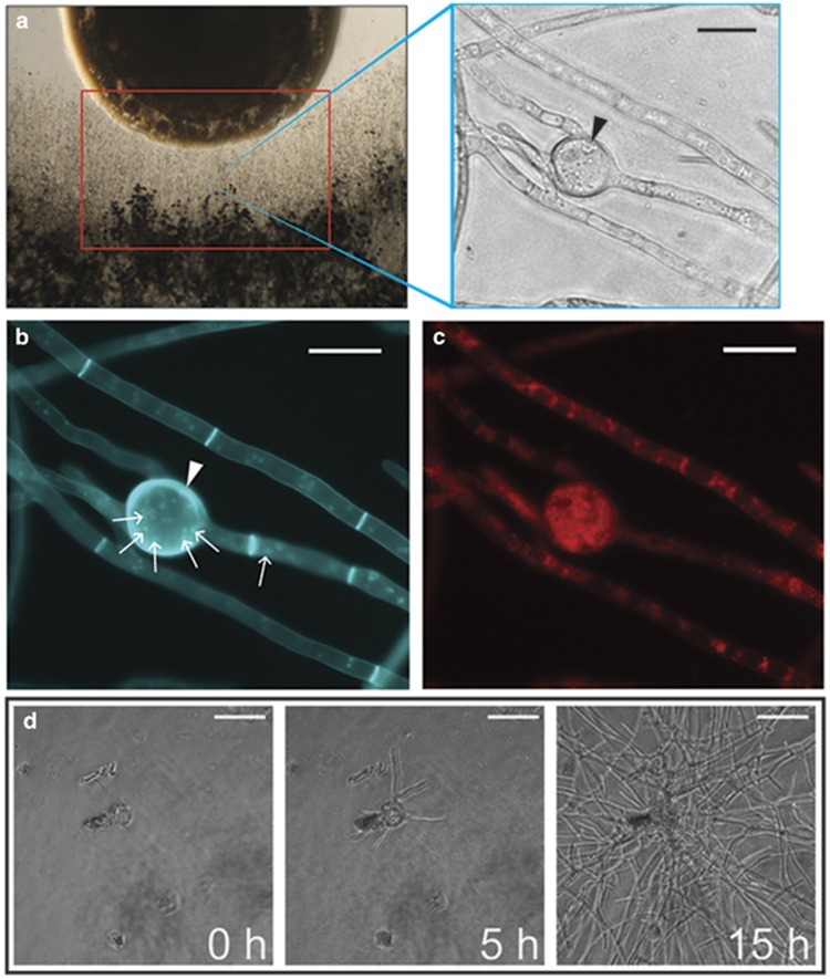
Examination of the interaction zone of the R. solanacearum/fungal coculture revealed fungal hypertrophy resembling chlamydospores produced by other fungi (a) left—coculture set-up of bacteria (top) and fungi (bottom). Red box indicates the ‘interaction zone' where many intercalated chlamydospore-like structures (right—black arrows) developed in response to coculture with R. solanacearum strain GMI1000. Control images of non-chlamydospore-inducing bacterial cocultures can bee seen in Supplementary Figure S1. Histological analysis of chlamydospores shows that they are (b) polynucleate (small white arrows), have thickened cell walls (white arrowheads) and (c) accumulate lipid bodies. (d) Time-lapse microscopy of isolated chlamydospores placed in fresh media shows these structures are independently viable, and they can germinate to form a new fungal colony. Complete time-lapse video can be viewed in Supplementary Video S1.

