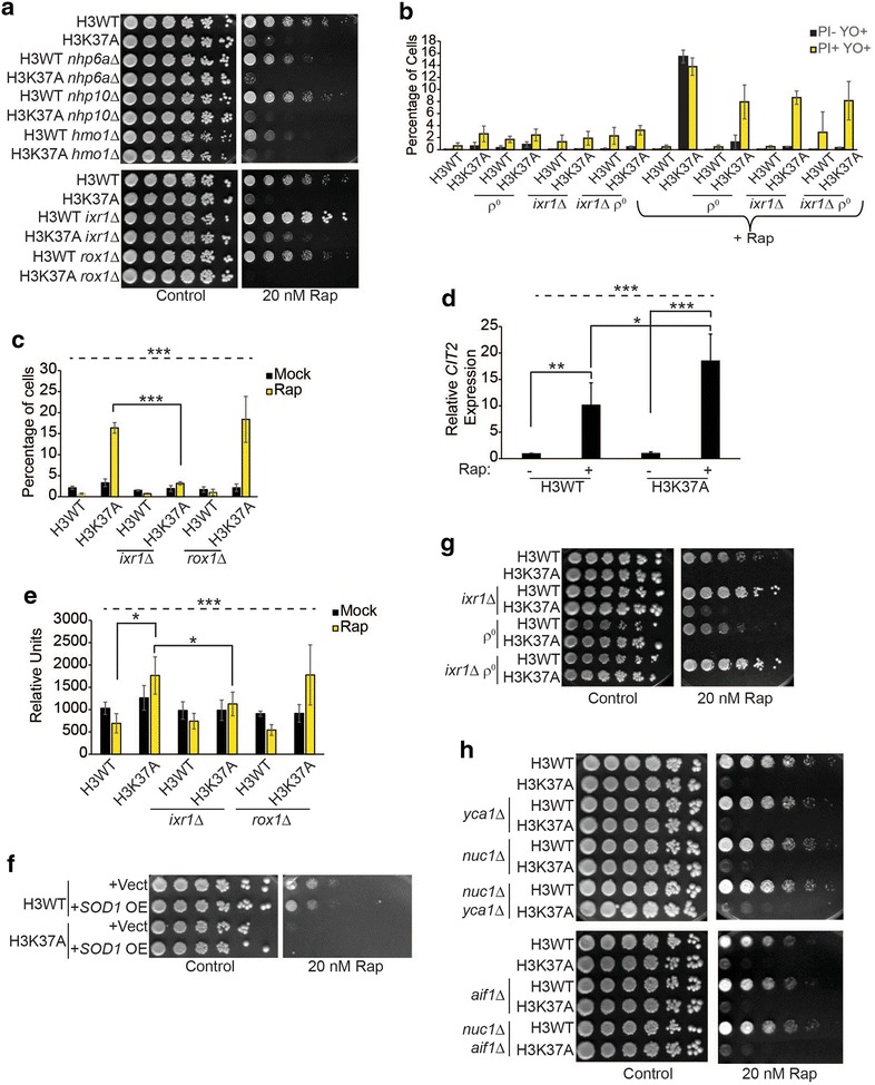
Fig. 4.

Histone H3 and TORC1 synergistically suppress both apoptosis and necrosis. a Spotting assay with H3WT, H3K37A, and the indicated HMG gene deletions. b Flow cytometry analysis of the indicated strains cultured to log phase and then mock treated or treated with 20 nM rapamycin for 5.5 h before staining with YO-PRO-1 and PI. Data are the average and SD of three independent experiments. c As in b except staining was performed only with YO-PRO-1 to solely detect apoptotic cells. d cDNA samples from H3WT and H3K37A mock or 20 nM rapamycin treated for 1 h were analyzed for CIT2 expression. Data are the average and SD of five independent experiments. e As in b except cells were stained with DHE. The average and SD of three independent experiments are presented. f Spotting assay on selective media (SC-Leu) with H3WT and H3K37A carrying either a control vector or an SOD1 high copy expression vector (OE). g Spotting assay with H3WT, H3K37A, and their derivatives lacking Ixr1 (ixr1∆), functional mitochondria (ρ°), or both. h As in a except genes encoding the indicated apoptotic effectors were deleted either individually or in combination. For all statistical analyses, one-way ANOVA was performed across all categories which is indicated by the dashed line, while the solid black lines indicate the specific pairwise comparisons which were analyzed by Student’s t test. *P < 0.05; **P < 0.01; ***P < 005
