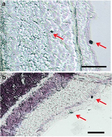
Fig. 4.

Expression of PK2 in the retinal ganglion cells of the mouse retina. PK2 immunolabeling was developed by DAB (3,3′-diaminobenzidine). a. Examples of two PK2-positive retinal ganglion cells, one nondisplaced and one displaced, are marked with arrows. b. PK2 DAB immunostaining with hematoxylin counterstaining. Examples of two PK2-positive retinal ganglion cells, one strongly and one more modestly stained, are marked with arrows. Scale bar, 20 μm
