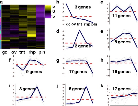
Fig. 7.

a-k Vision Heatmap for A. alata. Hierarchical clustering (EdgeR) and corresponding ten subcluster profiles for the 97 genes implicated in vision and the phototransduction pathway differentially expressed across A. alata medusa (gastric cirri, ovaries, tentacle (with pedalium base), rhopalium) and planulae samples. Intensity of color indicates expression levels for each of the ten hierarchical clusters (vertical access). Bright yellow patches correspond to the highest peaks for each k-mean subcluster profile. K-mean profiles (b-k) match the order of column names in a, representing the mean expression of gene clusters highly abundant in each sample (centroid demarcated by the solid line; zero indicated by the horizontal dashed red line). One bright yellow transcript clusters in the gastric cirri column correspond to a peak in plot b; one cluster in the ovaries column corresponds to plot c; three clusters in the tentacle column correspond to plots d, e and f; three bright yellow clusters in the rhopalium column correspond to peaks in plots h, i and j, and two less intense clusters correspond to peaks in plots g and k, and a slightly intense yellow gene cluster in the planulae column corresponds to the peak in plot k. The vertical colored bar on the left of the heatmap (a) indicates distinct patterns corresponding to the ten subcluster profiles (sc = subcluster number), for which the number of genes each comprises is indicated. Abbreviations: gc = gastric cirri, ov = ovaries, tnt = tentacle (and pedalium base), rhp = rhopalium, and pln = planulae. Gene annotations by subcluster provided in Additional file 8
