
Figure 1.
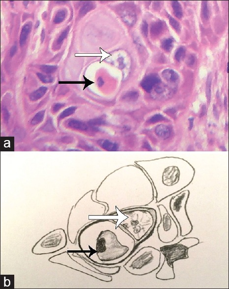
(a) Photomicrograph showing complete engulfment of apoptotic cell (black arrow) by oral squamous cell carcinoma tumor cell (white arrow) (H&E stain, ×400), (b) Hand-drawn illustration of the photomicrograph of 1a showing host cell (white arrow) and apoptotic cell (black arrow)