Figure 1.

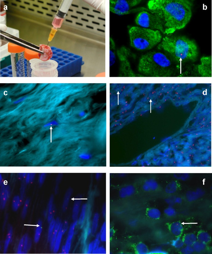
Male fetal microchimerism in female marrow, bone, lung and appendix. A section of rib was collected to obtain marrow (a), with adherent cell cultures (b) and sections of bone (c) analysed for the presence of the Y chromosome using XY-Fluorescence in situ hybridisation (FISH). A section of lung tumour (adenocarcinoma, d) was obtained at the same surgery from this cohort of postreproductive women. Sections of appendix were obtained from pregnant women undergoing clinically indicated appendicectomy (e, f). The X chromosomes in (d) and (e) are labelled with SpectrumOrange™, with the Y chromosomes labelled (d, e; arrows) with SpectrumGreen™. The Y chromosomes in (b, c and f) were identified using a Y FISH probe, labelled with SpectrumOrange™ (arrows, red signals). A male (red signal) CD3-positive (FITC, green) lymphocyte is shown in (f). Magnification ×100
