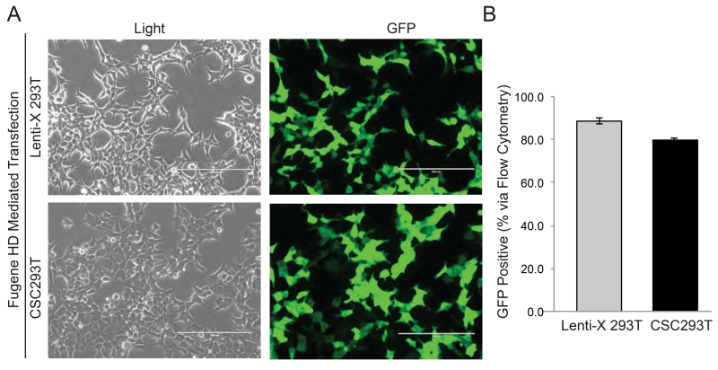
Figure 3. High Rates of Transfection in CSC293Ts.
(A) Representative images under light and fluorescent microscopy of Lenti-X 293Ts and CSC293Ts 24 hours post transfection with pGF1-CMV vector expressing GFP are shown with the white bar representing 200 μm. (B) Quantification of the percentage of GFP positive 293Ts 72 hours post transfection is shown.

