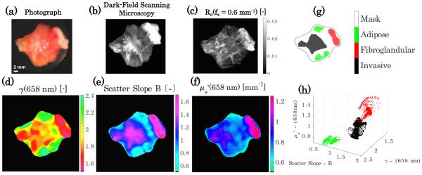
Fig. 6.
Spatial contexualization of scattering parameters for heterogeneous breast tissue. (a) Photograph of tissue, (b) dark field scanning microscopy, and (c) sub-diffusive calibrated reflectance image with f x = 0.6 mm−1 with no median filtering. (d)–(f ) Show scatter optical property maps of γ [−], scatter slope B [−], and [mm−1], respectively. (g) Regions of interest corresponding to areas of localized tissue diagnoses and (h) clustering of the scatter properties for each tissue diagnosis.

