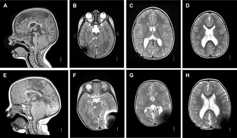
Figure 1. MM-associated hydrocephalus. A–D: Classic Chiari II malformation.

Sagittal T1 MRI image (A) demonstrating classic features of a Chiari II malformation including elongated pons and downwardly displaced medulla, tectal beaking, small posterior fossa with vertically oriented tentorium, with cerebellar tonsillar ectopia below the foramen magnum line. Axial l T2 images (B–D). Note patency of aqueduct in B (arrow). E–H: Chiari II with aqueductal compression. Sagittal T1 image (A) demonstrating similar anatomic configuration as A, but with more prominent posterior fossa crowding and aqueductal compression. Axial T2 images (F–H). Note absence of patent aqueduct in F (arrow).
