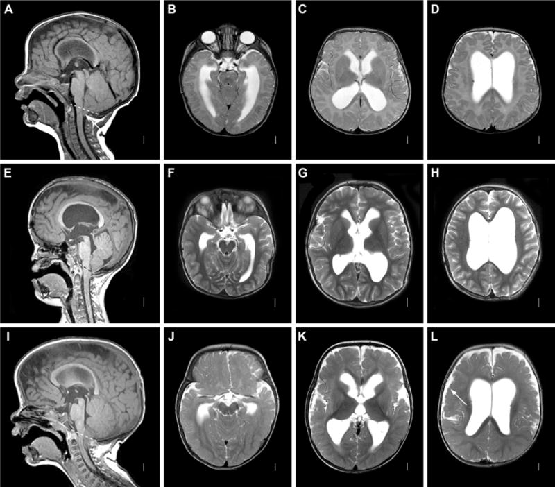
Figure 3. Posterior fossa crowding. A–D: Chiari I malformation.

Axial T1 MRI image (A) showing open aqueduct and relatively large-appearing cerebellum, with herniation of tonsils below foramen magnum (dashed line). Axial T2 images (B–D) showing moderate dilatation of lateral ventricles. E–H: Pfeiffer Syndrome with multi-suture synostosis. Sagittal T2 images (E) showing midface retrusion, widely patent aqueduct and small, crowded posterior fossa with tonsillar herniation below the foramen magnum (dashed line) in patient with a confirmed FGFR2 mutation. Axial T2 images (F–H) showing moderate ventricular dilatation. I–L: MPPH syndrome. Sagittal T1 image (I) showing widely patent aqueduct with tonsillar herniation through foramen magnum (dashed line). Axial T2 image showing moderate ventricular dilation (J–L) and extensive bilateral perisylvian polymicrogyria (L, arrow).
