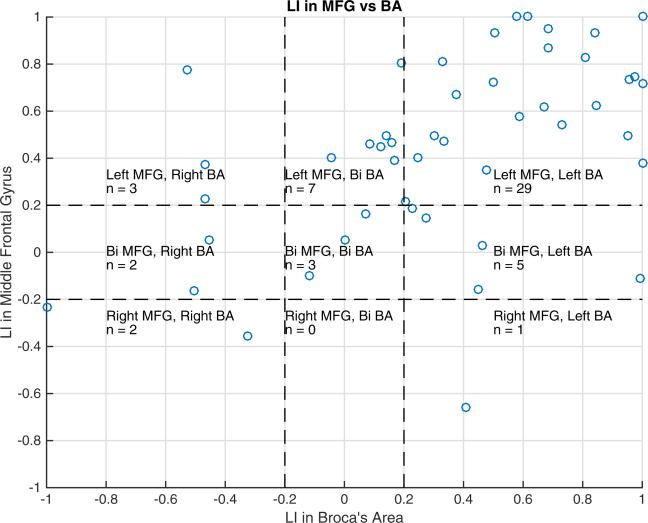
Fig. 2.
Scatterplot of LIs in MFG versus BA. There is a positive correlation between LIs in MFG and BA (Spearman r = 0.62, p < 0.001). Of the 52 subjects, 29 (56 %) have left laterality in both MFG and BA, 7 (13 %) have left laterality in MFG and bilaterality in BA, while 5 (10 %) have bilaterality in MFG and left laterality in BA. The remainder of the subjects includes three (6 %) with bilaterality in MFG and BA, three (6 %) with left laterality in MFG and right laterality in BA, one (2 %) with right laterality in MFG and left laterality in BA, two (4 %) with bilaterality in MFG and right laterality in BA, and two (4 %) with right laterality in both MFG and BA.
Bi = bilateral

