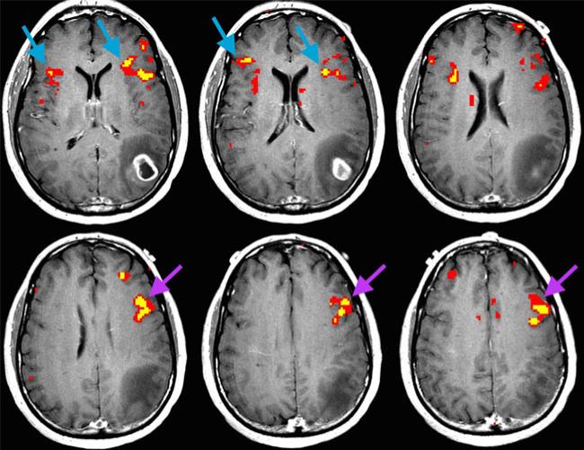
Fig. 3.
Axial fMRI cross section of a 50-year-old man with glioblastoma in the left parietal lobe (subject no. 40). In this case, language fMRI revealed bilaterality in BA (blue arrows) and left laterality in MFG (purple arrows). MFG may be helpful in such a case where BA is equivocal for language hemispheric dominance

