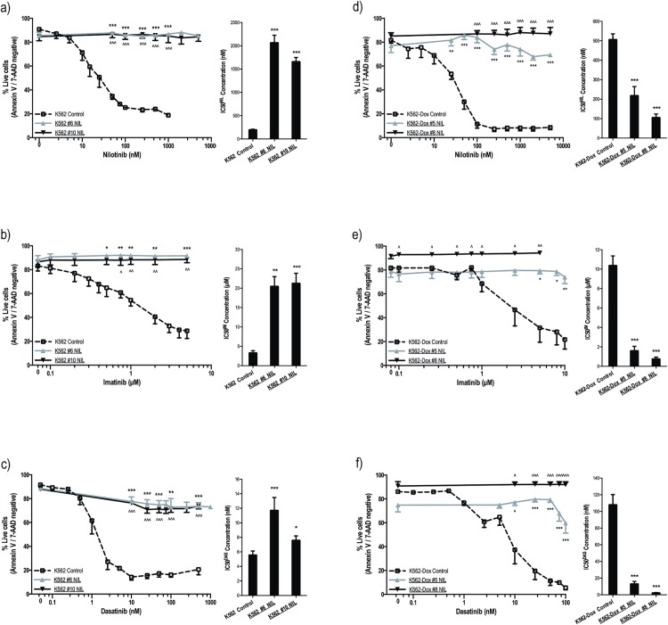
Fig 1. K562 and K562-Dox cells cultured long term in nilotinib demonstrate cross-resistance to imatinib and dasatinib.
(a-c) K562 and (d-f) K562-Dox cells were incubated for 3 days with the indicated concentrations of (a,d) nilotinib, (b,e) imatinib or (c,f) dasatinib. Cell viability was determined by Annexin V/7-aminoactinomycin D staining in at least three independent experiments and expressed as %live cells (line graphs). p-CRKL dependent IC50 (dose of TKI required to reduce p-CRKL levels by 50%) was calculated by western blot in at least three independent experiments. Average IC50 values of the corresponding densitometry analyses are denoted in the column graphs. Representative western blots are shown in S1 Fig. Cell viability statistical analyses compared % live cells at common TKI concentrations; western blot statistical analyses compared resistant cells to corresponding controls. Analyses were performed using unpaired Student’s t-test (Welch’s correction was applied for data groups with unequal SD) or Mann-Whitney Rank Sum test. Statistically significant p-values are denoted by carets (^) or asterisks (* p<0.05; ** p<0.01; *** p<0.001). Error bars represent SEM. NIL = nilotinib.

