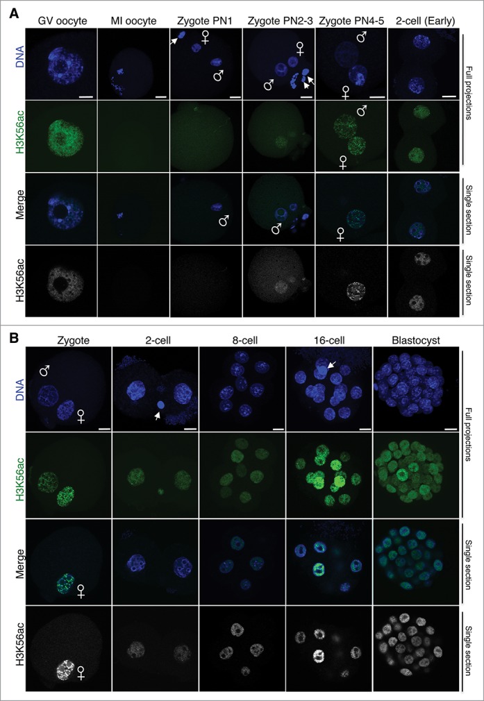
Figure 3.

H3K56ac is first detected at the mid-zygote stage after fertilization and persists throughout pre-implantation development. (A) Analysis of H3K56ac immediately after fertilization and until the early 2-cell stage. Freshly collected mouse embryos from natural matings at the indicated stages were stained with an H3K56ac antibody (green) and with DAPI (blue). Full projections of Z-sections acquired every 1 μm (top 2 parts) and middle, merge section (third part) are shown. The gray scale view of the green (H3K56ac) channel of the merge image confocal section is shown at the bottom. Male and female pronuclei are indicated, and the polar body, where visible, is demarcated by an arrow. The PN classification was done as described by Adenot and colleagues.20 At least 10 embryos were analyzed per stage, performed in 2 independent experiments. Scale bar is 12 μm. (B) Dynamics of H3K56ac throughout cleave stages. Embryos were collected at the indicated stages, fixed, and processed for immunostaining using an H3K56ac. As in A, top and second panels are maximal projections of confocal Z-sections acquired every 1 μm (cleavage stages) or 2 μm (blastocyst) of DAPI (blue) or H3K56ac (green) channels. The merge image shows a middle confocal section of the same embryos and the bottom panel is the gray scale of the green (H3K56ac) channel. At least 14 embryos were analyzed per stage, performed in 2 independent experiments. Scale bar is 12 μm.
